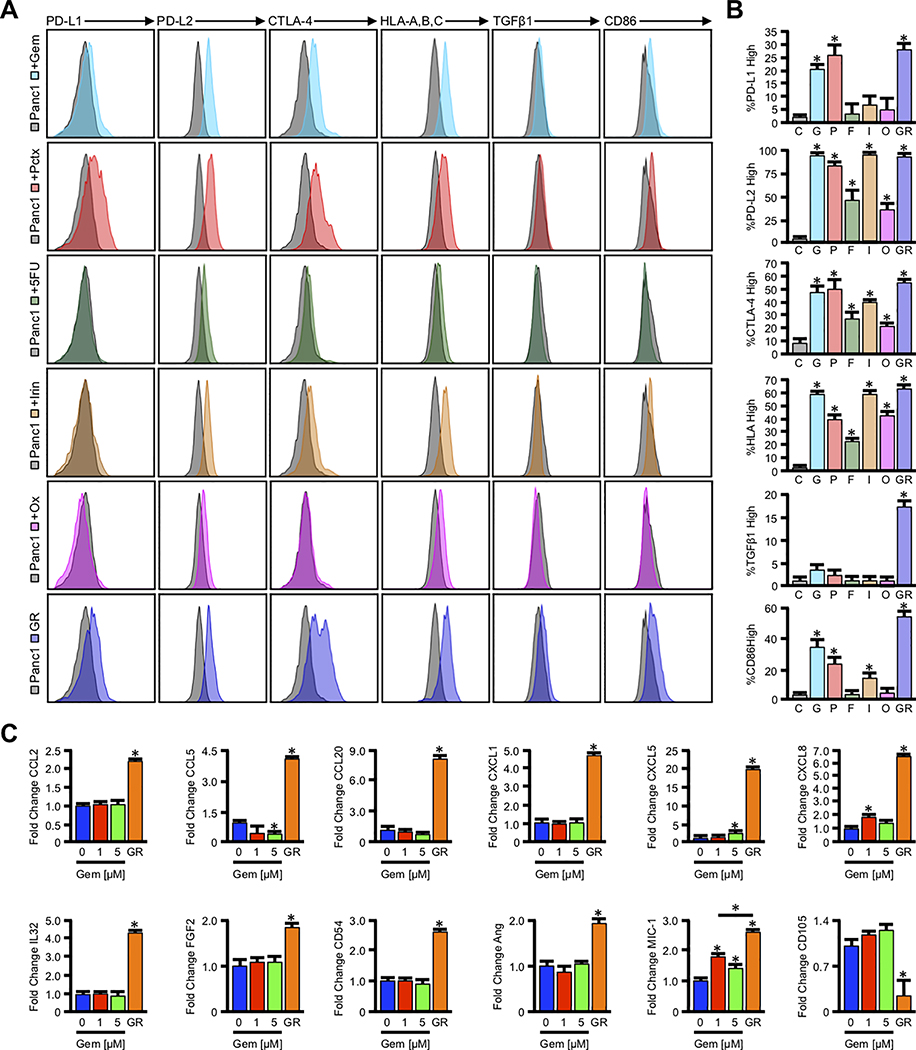
Figure 3. Cytotoxic chemotherapy alters tumor cell immunogenicity in vitro.
(A) Panc1 cells were incubated with the known IC50 of the first line chemotherapy agents Gemcitabine (Gem, 1μM), Paclitaxel (Pct, IC50 = 100nM), 5-Fluorouracil (5-FU, 2.5μM), Irinotecan (Irin, 2.5μM), and Oxaliplatin (Ox, 2.5μM), after which the expression of both surface and intracellular immmunomodulatory proteins was evaluated by flow cytometry. (B) Using unstained Panc1 cells, we identified the number of cells positive for each antigen in vehicle treated control (C) cells. We then determined the percent of the parent population for Gemcitabine (G), Paclitaxel (P), 5-Fluorouracil (F), Irinotecan (I), and Oxaliplatin (O) treated Panc1 cells, or Panc1-GR (GR) cells with expression above the geometric mean for control cells (*p < 0.05). (C) Panc1 cells were incubated with 0, 1, or 5μM Gemcitabine over a 48-hour period, and Panc1-GR cells were grown in 10μM Gemcitabine for several passages. Cells were incubated with a protein transport inhibitor for one hour, lysed, and 200μg of total cell lysate was evaluated by a high throughput proteome profiler array (ARY022B). Pixel density was evaluated using ImageJ, and samples normalized to the mean intensity of the reference spots for each blot minus the background density. Composite normalized values for all experimental groups were divided by those for untreated Panc1 cells, and are presented as fold change plus standard deviation (*p < 0.05).

