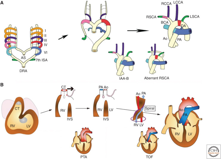
Figure 2.
Development and anomalies of the aortic arch (AA) system and the cardiac outflow tract (OFT). (A) Remodeling of bilaterally symmetric pharyngeal arch arteries (PAAs) into the AA system is shown in a color-coordinated fashion. Interrupted AA type B (IAA-B) results from abnormal regression of the left fourth PAA. Aberrant right subclavian artery (RSCA) results from abnormal regression of the right fourth PAA. (B) Developmental steps of the cardiac OFT, including leftward movement (bold arrow) of the conotruncus (CT) and septation of the CT into the aorta (Ao) and the pulmonary artery (PA) in a spiral fashion are shown. Persistent truncus arteriosus (PTA) resulting from a failure of septation of the CT. Tetralogy of Fallot (TOF) resulting from malalignment of the Ao to the left ventricle (LV). (I–VI) first to sixth PAA, (AS) aortic sac, (BCA) brachiocephalic artery, (DA) ductus arteriosus, (DRA) dorsal artery, (ISA) intersegmental artery, (IVS) interventricular septum, (LCCA) left common carotid artery, (LSCA) left subclavian artery, (RCCA) right common carotid artery, (RSCA) right subclavian artery, (RV) right ventricle.

