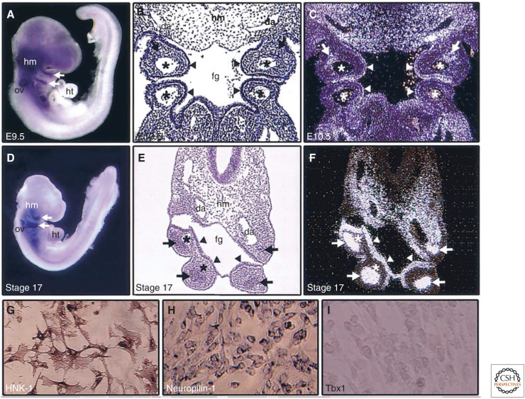
Figure 5.
Expression patterns of Tbx1. Mouse (A–C) and chick (D–F) embryos and primary neural crest culture from chick embryos (G–I) are shown. RNA in situ hybridizations for whole-mount (A,D), coronal section (B,C), and transverse section (E,F) show Tbx1 expression (blue or white signals). In the pharyngeal arches, Tbx1 is expressed in mesodermal core (arrows) and endodermal epithelium (arrow heads), excluding the neural crest–derived mesenchyme surrounding the mesodermal core. Tbx1 is also expressed in head mesenchyme (hm) and otic vesicle (ov). B and E are bright field images of C and F, respectively. Asterisks indicate pharyngeal arch arteries. In chick primary neural crest culture cells, HNK-1 (G) and neuropilin-1 (H) are detected by immunocytochemistry and mRNA in situ hybridization, respectively, but Tbx1 is not detectable by mRNA in situ hybridization (I). (fg) foregut, (da) dorsal aorta, (ht) heart (from Yamagishi 2002, with permission).

