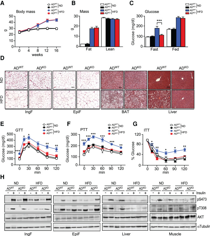
Figure 1.
HFD-induced metabolic syndrome is suppressed in mice with JNK deficiency in adipocytes. (A) Whole body mass of ND-fed and HFD-fed (16 wk) ADWT and ADKO mice was measured (mean ± SEM; n = 8∼9). (B) The fat mass and lean mass of ND-fed and HFD-fed (16 wk) mice were measured using 1H-MRS (mean ± SEM; n = 8∼9). (C) The blood glucose concentration of ND-fed and HFD-fed (16 wk) mice was measured (mean ± SEM; n = 8∼9). (***) P < 0.001. (D) Sections prepared from ND-fed and HFD-fed (16 wk) ADWT and ADKO mice were stained with H&E. Scale bar, 100 µm. (E–G) Tolerance tests for glucose (GTT), pyruvate (PTT), and insulin (ITT) were performed using ND-fed and HFD-fed (16 wk) mice (mean ± SEM; n = 7∼9). (*) P < 0.05; (**) P < 0.01; (***) P < 0.001. (H) ND-fed and HFD-fed (16 wk) mice were treated with insulin (15 min). Immunoblot analysis was performed with extracts prepared from adipose tissue (IngF and EpiF), liver, and gastrocnemius muscle using antibodies to phospho-AKT, AKT, and αTubulin.

