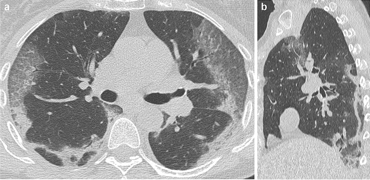
Fig. 2.
61-year old woman with confirmed COVID-19 pneumonia. At hospital admission, HRCT images on axial (a) and sagittal (b) planes showed bilateral, peripheral GGO)and band-like opacities with a perilobular distribution, resembling an OP pattern. OP pattern was deemed present by all readers. After 5 days, the patient developed respiratory failure [Italian Society of Emergency Medicine (SIMEU) phenotype IV disease]

