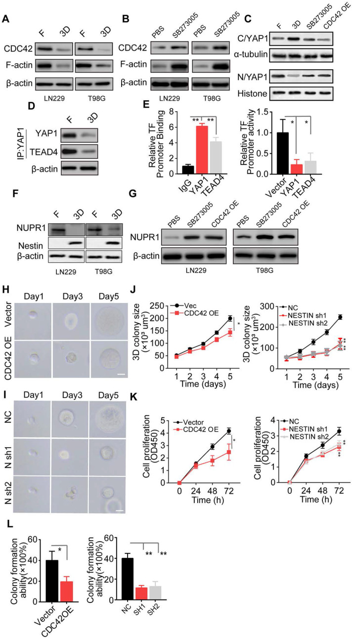
Figure 4.
The integrin αvβ3/CDC42/F-actin/YAP/NUPR1/Nestin signaling pathway is activated in collagen/FN-cultured glioma cell (A) Expression of CDC42, F-actin, and β-actin in LN229 cells and T98G cells cultured in a flask or 3D Collagen/FN gel. (B) Expression of CDC42, F-actin, and β-actin in LN229 cells and T98G cells cultured in 3D Collagen/FN gel treated with PBS or SB273005 (5 nM, 24). (C) Cell fraction of cytosol (C) and nucleus (N) was analyzed by Western blotting. Expression of YAP1 in LN229 cells or LN229 CDC42 OE cells cultured in a flask or 3D Collagen/FN gel and treated with PBS or SB273005 (5 nM, 24 h). (D) Western blotting analysis of proteins immunoprecipitated (IP) with anti-YAP1 from LN229 cells cultured in a flask or 3D Collagen/FN gel for 48 h. (E) ChIP analysis of YAP1 or TEAD4 binding to the NUPR1 promoter in LN229 cells. YAP1 and TEAD4 increased the luciferase activity of the NUPR1 promoter in LN229 cells. (F) Expression of NUPR1 and Nestin in LN229 cells and T98G cells cultured in a flask or 3D Collagen/FN gel. (G) Expression of NUPR1in LN229 and T98G cells cultured in 3D Collagen/FN gel, treated with PBS or SB273005 (5 nM, 24 h) and transfected with CDC42 OE lentivirus. (H) Representative photographs of LN229-NC cells and LN229-CDC42 OE cells cultured in 3D Collagen/FN gel. Scale bar represents 20 µm (I) Representative photographs of LN229-NC cells and LN229-Nestin shRNA cultured in 3D Collagen/FN gel. Scale bar represents 20 µm (J) Quantification of colony sizes in (H) and (I) (K) Proliferation of 3D collagen/FN pre-cultured LN229-vec cells, LN229-CDC42 OE cells, LN229-NC cells, and LN229-Nestin shRNA cells (L) Colony formation of 3D collagen/FN pre-cultured LN229-vec cells, LN229-CDC42 OE cells, LN229-NC cells, and LN229-Nestin shRNA cells. Mean ± SEM, n.s, no significant difference, *p < 0.05, **p < 0.01.

