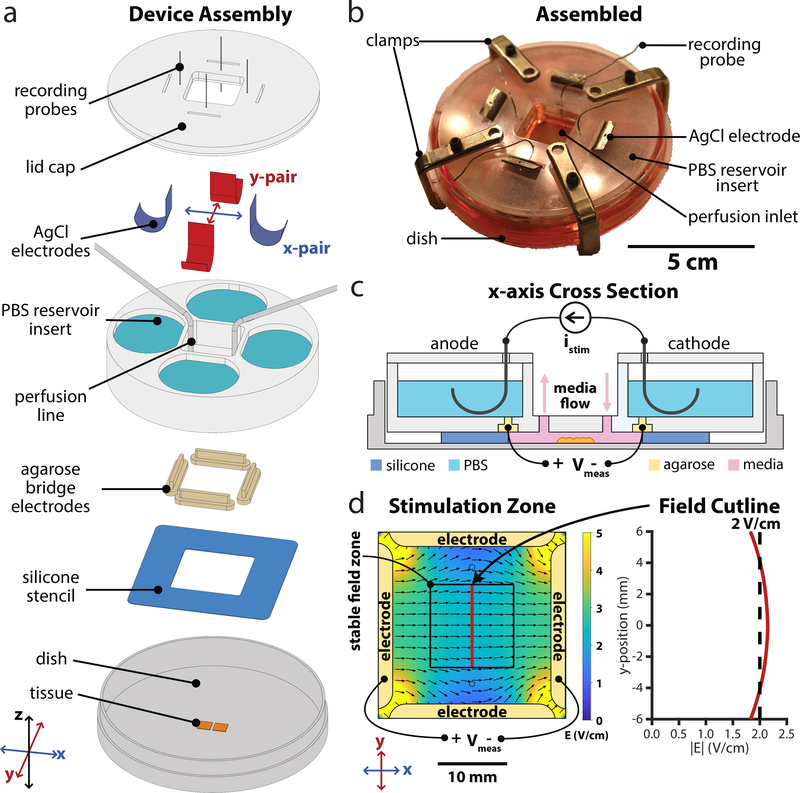
Figure 1.
Design of next-gen electro-bioreactor for multi-axis migration control; SCHEEPDOG: Spatiotemporal Cellular HErding with Electrochemical Potentials to Dynamically Orient Galvanotaxis, a bioreactor for two-dimensional, bioelectric control over collective migration. (a) Assembly of the SCHEEPDOG bioreactor within a Petri dish. A silicone stencil defines a stimulation zone in the center of the dish, and tissues are patterned within this region. Four agarose bridges held inside a laser-cut PBS reservoir insert are then pressed against the stencil to fully enclose the stimulation zone. Ag/AgCl electrodes and perfusion lines are added, then a lid is clamped to the assembly and recording probes are inserted into each agarose bridge electrode. (b) Photograph of a fully assembled device. (c) Sectional schematic of one axis of SCHEEPDOG. Each axis features one anode/cathode pair connected to a computer-controlled current supply which provides istim. A digital oscilloscope records channel voltage Vmeas to enable closed-loop control. (d) Numeric simulation of the field generated by the device when the horizontal electrode pair is stimulated. The simulated field value is indicated with a red line showing a stable field zone in the center of the device. Related to Supplemental Figs. 1,2.

