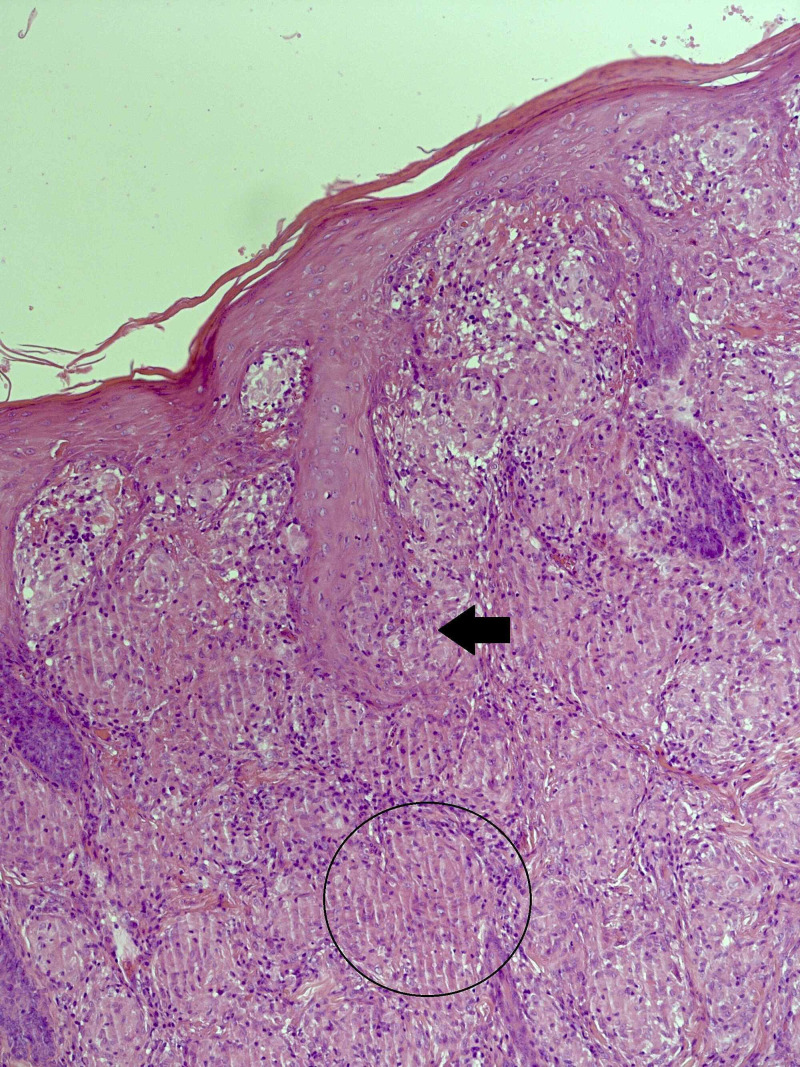
Figure 2. A section of skin stained with H&E. The dermis is packed with relatively well-defined non-necrotizing granulomata, some of which are surrounded by a loose cuff of lymphocytes (circled area). The skin adnexae are involved and disrupted by the granulomata (black arrow).
H&E, hematoxylin and eosin

