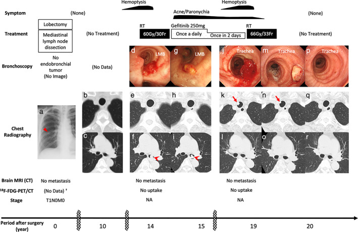
Figure 1.

Clinical course. Initial chest X‐ray imaging (a, triangle) revealed a primary lung adenocarcinoma. Ten years after complete resection, there were no abnormal CT findings in the trachea (b) or LMB (c). Fourteen years after surgery, bronchoscopy (d) and CT (f, arrowhead) demonstrated the first EEM localized in the LMB, with no tumor in the trachea (e). Gefitinib was initiated after irradiation therapy (60 Gy/30 Fr). Because of these treatments, bronchoscopy (g) and CT showed no tumors in the trachea (h), and LMB lesions (i, arrowhead) were diminished and eventually disappeared (l, o, r). Nineteen years after surgery, the second EEM was confirmed by bronchoscopy (j) and CT (k, arrow) in the trachea, and RT was administered (66 Gy/33 Fr). One month later, bronchoscopy (m) and CT (n, arrow) revealed that the EEM had shrunk. One year after the last treatment, bronchoscopy (p) and CT (q) showed no endotracheal lesions. 18F‐FDG‐PET/CT and brain MRI confirmed no further metastasis during this period. †Instead of 18F‐FDG‐PET/CT, CT with contrast and bone scintigraphy was performed, and no metastasis was found. CT, computed tomography; EEM, endotracheal/endobronchial metastasis; FDG, fluorodeoxyglucose; Fr, fractions; LMB, left main bronchus; MRI, magnetic resonance imaging; PET, positron emission tomography; RT, radiation therapy; NA, not applicable.
