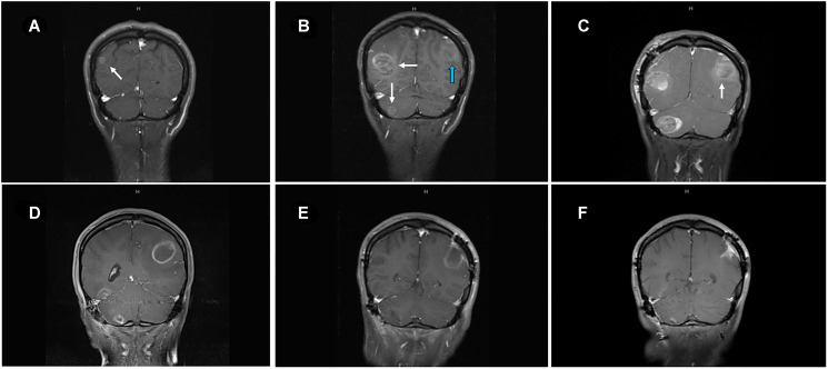
Figure 2.
MRI of brain of metastatic lesions showing (A) coronal section demonstrating right parieto-occipital lesion at the start of ipilimumab-nivolumab (arrow). (B) coronal image showing right parieto-occipital and right cerebellar lesions (white arrows) and left posterior parietal lesion (blue arrow) after three cycles of ipilimumab-nivolumab. (C) Post-surgical resection of two symptomatic lesions- right parieto-occipital and right cerebellar lesions with persistent left posterior parietal lesion (arrow). (D) Enlarging left posterior parietal lesion post TVEC and pembrolizumab. (E) Surgical cavity after resection of left posterior parietal lesion and one month after TMZ treatment. (F) Collapsing surgical cavity of left posterior parietal lesion resection with no evidence of recurrent disease in the brain after 19 months of temozolomide initiation.

