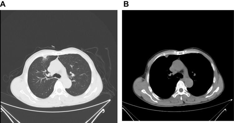
Figure 3.
Chest enhanced CT after neoadjuvant immunotherapy: (A) The area of the anterior segment of the right upper lobe of the lung was significantly smaller than that of the anterior, with a maximum cross-section of about 11 mm × 7 mm. (B) The mediastinal and right hilar swollen lymph nodes were smaller than before, and the adjacent pulmonary vessels were more compressed Before remission.

