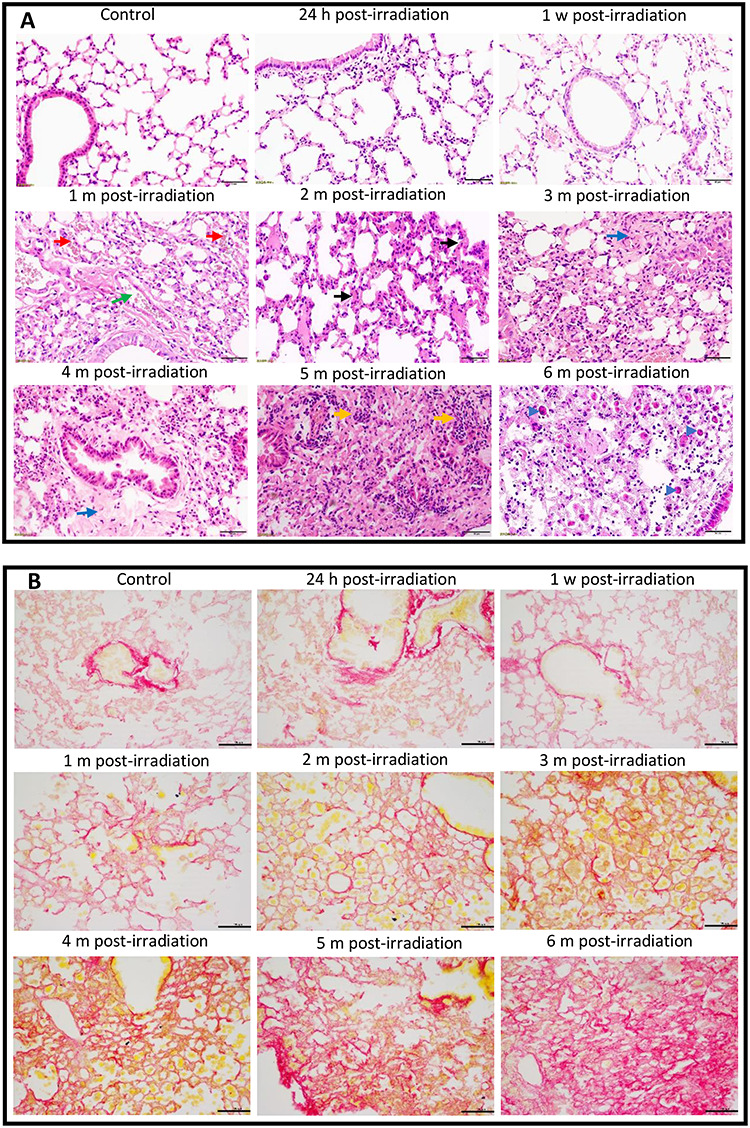
Fig. 1.

Histopathological changes in the lungs of mice after thoracic irradiation. Female C57BL/6 J mice received 20 Gy of X-rays in a single dose. The mice were euthanized at 24 h, 1 week, 1, 2, 3, 4, 5 or 6 months post-irradiation. The mice in the corresponding control groups were sacrificed at the same time points. (A) Photomicrographs of lung tissues stained with H&E. Green arrow, a dilated vessel; red arrows, congestive capillaries; black arrows, thickened septa; blue arrows, fibrotic tissue; yellow arrows, inflammatory cells; blue arrowheads, macrophages, mononucleolar cells or foam cells. (B) Images of lung tissues stained with Picro-Sirius Red. Collagen stains red. Scale bar = 50 μm. H, hour; w, week; m, month.
