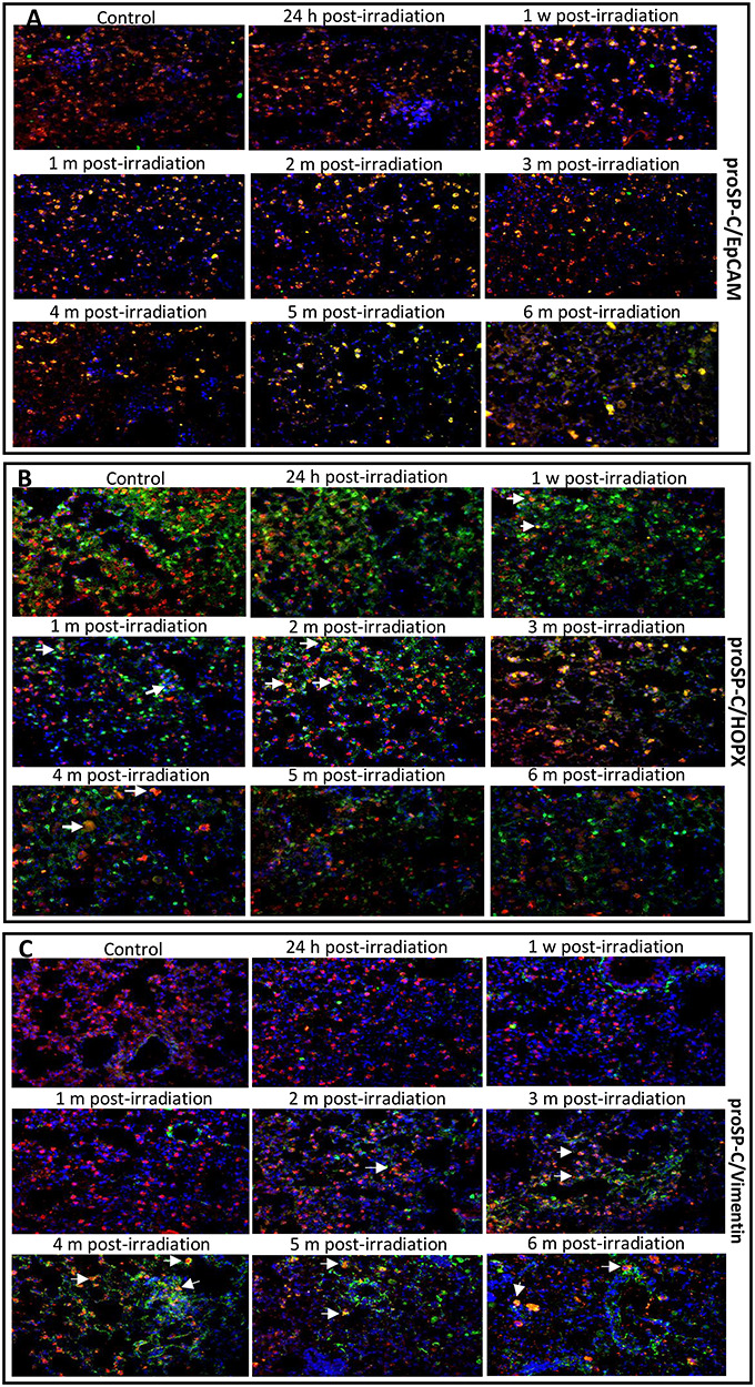
Fig. 2.

The merged images of the coexpression of proSP-C (AEC II cell marker) with EpCAM (epithelial stem cell marker) (A), HOPX (AEC I cell marker) (B), or vimentin (mesenchymal cell marker) (C) in the lungs of mice treated with IR and control-treated mice. Double immunofluorescence staining was used to detect protein expression. In the representative photomicrographs, the color of the pro-SP-C protein is red, the EpCAM, HOPX and vimentin proteins are green, and the nuclei are blue. The yellow fluorescence in AEC II is due to the overlap of red and green (arrows). Coexpression of EpCAM/proSP-C, HOPX/proSP-C or vimentin/proSP-C in the lung tissues was used to track AEC II dynamic phenotypes at different phases of injury after irradiation. Magnification: 400x. h, hour; w, week; m, month.
