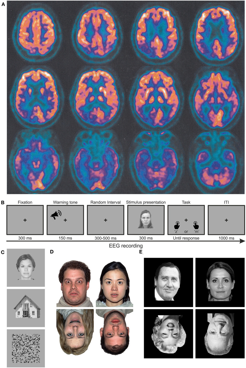
Figure 1.
Material and methods. (A) Axial PET images of patient MCG at the time of diagnosis, highlighting significant hypometabolism in the bilateral inferior temporal poles and in the occipito-parieto-temporal cortex, lateralized to the right hemisphere. Images are shown according to the radiological convention (right is left and vice versa). (B) Trial structure of the performed discrimination tasks. In experiment 1, participants had to discriminate whether the stimulus was meaningful or not. In experiments 2 and 3, participants had to discriminate whether the stimulus was upright or inverted. (C–E) Examples of stimuli employed in experiments 1 (faces, houses, and scrambled images), 2 (upright and inverted faces with neutral or fearful expressions, respectively), and 3 (upright and inverted faces for famous or unfamiliar faces).

