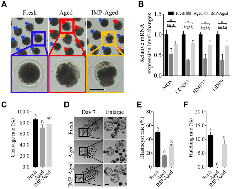
FIGURE 1.
IMP supplementation enhanced the development of porcine parthenogenetic embryos derived from aged oocytes. (A) Representative images of oocytes in fresh, aged, and IMP-treated aged groups. Normal/abnormal cytoplasm/spontaneous parthenogenetic activated oocytes were labeled with blue/red/orange arrows. Scale bar = 50 μm. (B) Differential gene expression in fresh, aged, and IMP-treated aged oocytes. Gene expression was detected in oocytes after 24 h of aging. R = 3. *P < 0.05; **P < 0.01. (C) Cleavage rate of parthenogenetic activated embryos derived from fresh (N = 199), aged (N = 216), and IMP-treated aged (N = 218) group. R = 4. Significant differences are represented by different capital letters (P < 0.01). (D) Representative images of parthenogenetic embryo development on day 7 derived from fresh, aged, and IMP-treated aged groups. Scale bar = 100 μm. (E) Blastocyst formation rate on day 7 in the fresh, aged, and IMP-treated aged groups. Significant differences are represented by different capital letters (P < 0.01). (F) Hatching rate on day 7 in the fresh, aged, and IMP-treated aged groups. No hatched blastocysts were observed in aged group. Significant differences are represented by different lower-case (P < 0.05) and capital (P < 0.01) letters.

