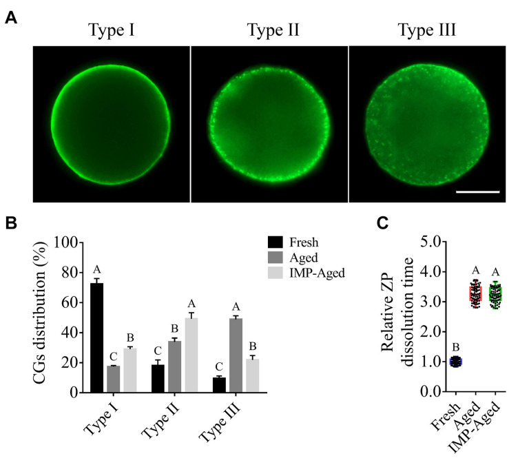
FIGURE 2.
Effects of IMP on the distribution of cortical granule and ZP hardness in aged oocytes with/without IMP treatment. (A) Representative images of different types of cortical granule distribution. Scale bar = 25 μm. (B) The proportion of different types of oocytes in the fresh (N = 105), aged (N = 98), and IMP-treated aged groups (N = 100). R = 3. Significant differences are represented by different capital letters (P < 0.01). (C) Relative dissolution time of ZP in fresh (N = 75), aged (N = 75), and IMP-treated aged (N = 75) oocytes. No significant difference was observed in dissolution time of ZP between aged and IMP-treated aged oocytes, which suggested no significant difference in ZP hardness with or without IMP treatment. R = 3. Significant differences are represented by different capital letters (P < 0.01).

