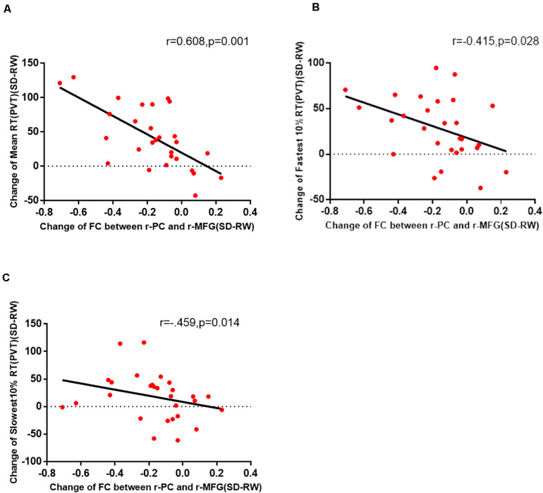
FIGURE 4.
The change in functional connectivity between the right PC and the right MFG was significantly correlated with the mean RT (A), fastest 10% RT (B), and slowest 10% RT (C) of PVT. PC, precuneus; MFG, right middle frontal gyrus; PVT, psychomotor vigilance test; SD, sleep deprivation; RW, rested wakefulness; RT, response time.

