
Figure 2.
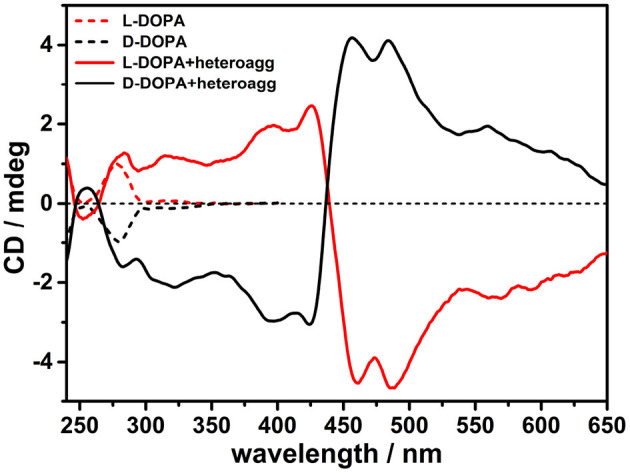
Circular dichroism (CD) spectra in phosphate buffered saline (PBS) buffer (pH = 7.4) of porphyrin hetero-aggregates [5,10,15,20-tetrakis(4-N-methylpyridyl)porphyrin tetrachloride salt (H2T4) = 4 μM, copper(II) 5,10,15,20-tetrakis(4-sulfonatophenyl)porphyrin tetrasodium salt (CuTPPS) = 4 μM] in the presence of L-3,4-dihydroxyphenylalanine (L-DOPA; red solid curve) and D-3,4-dihydroxyphenylalanine (D-DOPA; black solid curve) as prepared. The CD spectra for dihydroxyphenylalanine (DOPA) alone in PBS buffer are graphed in red dashed curve for L-enantiomer and in black dashed curve for D-enantiomer. In all samples, the concentration of DOPA was 0.5 mM.