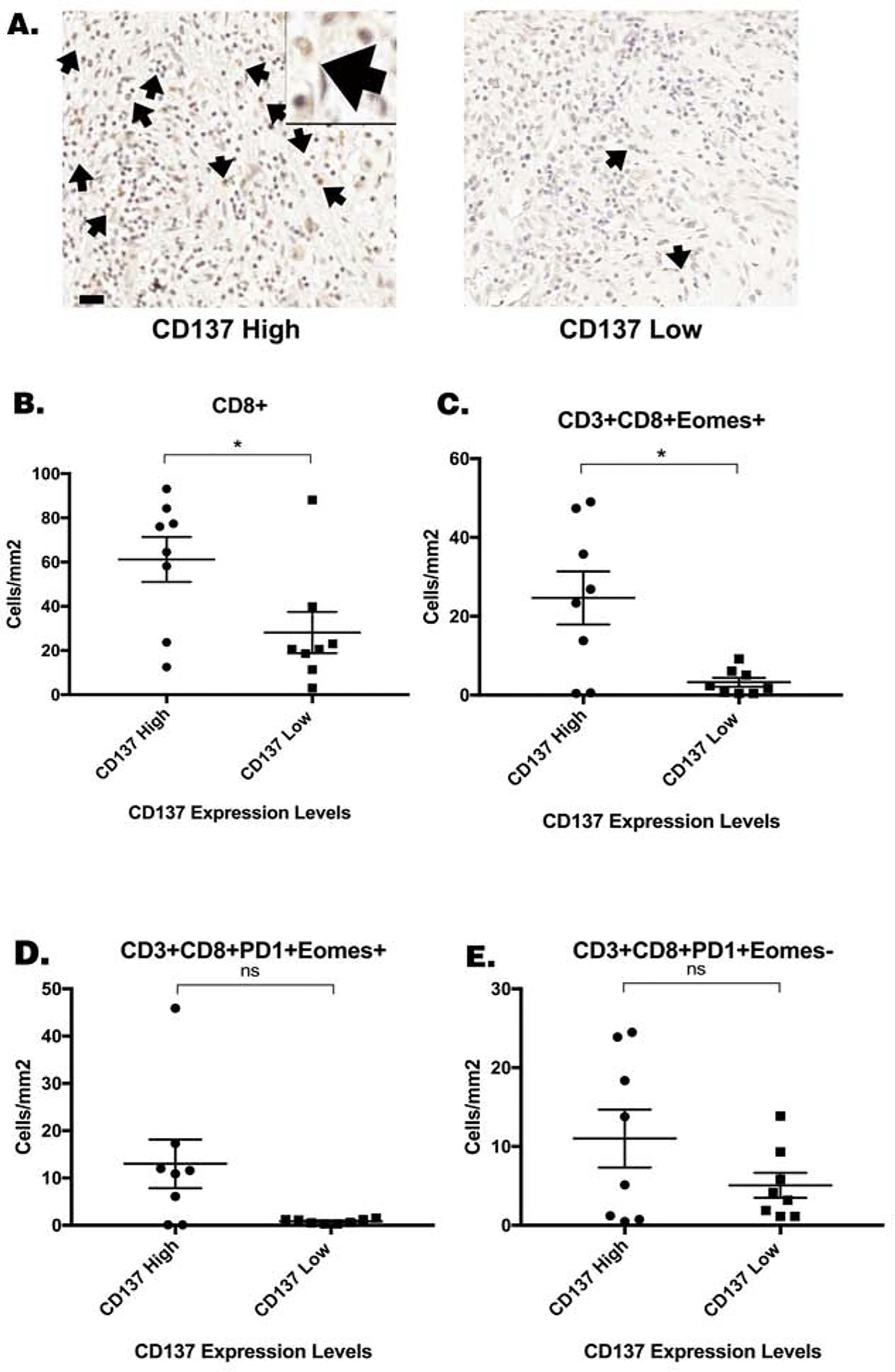
Figure 2. High CD137 expression in human PDAC treated with vaccine therapy was associated with increased CD8+ T cell density.

(A.) Sixteen formalin-fixed paraffin-embedded human PDAC specimens were obtained following surgical resection two weeks after one neoadjuvant dose of GVAX were analyzed by multiplex immunohistochemistry as previously described [18]. Immunohistochemistry staining of CD137 was performed on slides from each of these 16 FFPE blocks. CD137+ cells was quantified and the median density of CD137+ cells (81.6 cells/cm2) was used as the cutoff to group high CD137+ cell density (greater than 81.6 cells/cm2) and low CD137+ cell density (less than 81.6 cells/cm2) patients. Representative cases with high CD137+ cell density and low CD137+ cell density, respectively, were shown. Representative CD137+ cells were indicated by arrows. One representative CD137+ cell were shown in a higher power field. Scale bar, 20 μm. (B.) The correlation between the density of CD8+ T cells as previously determined[18] by multiplex immunohistochemistry and the density of CD137+ cells in the PDAC tumors was analyzed in this study. The densities of (C.) CD3+CD8+Eomes+ cells (D.) CD3+CD8+PD-1+Eomes+ cells, and (E.) CD3+CD8+PD-1+Eomes− T cells in the tumor area of PDAC patients as previously determined[18] by multiplex immunohistochemistry, respectively, were correlated with the density of CD137+ cells. Unpaired t tests were done. * p<0.05. ns, not significant.
