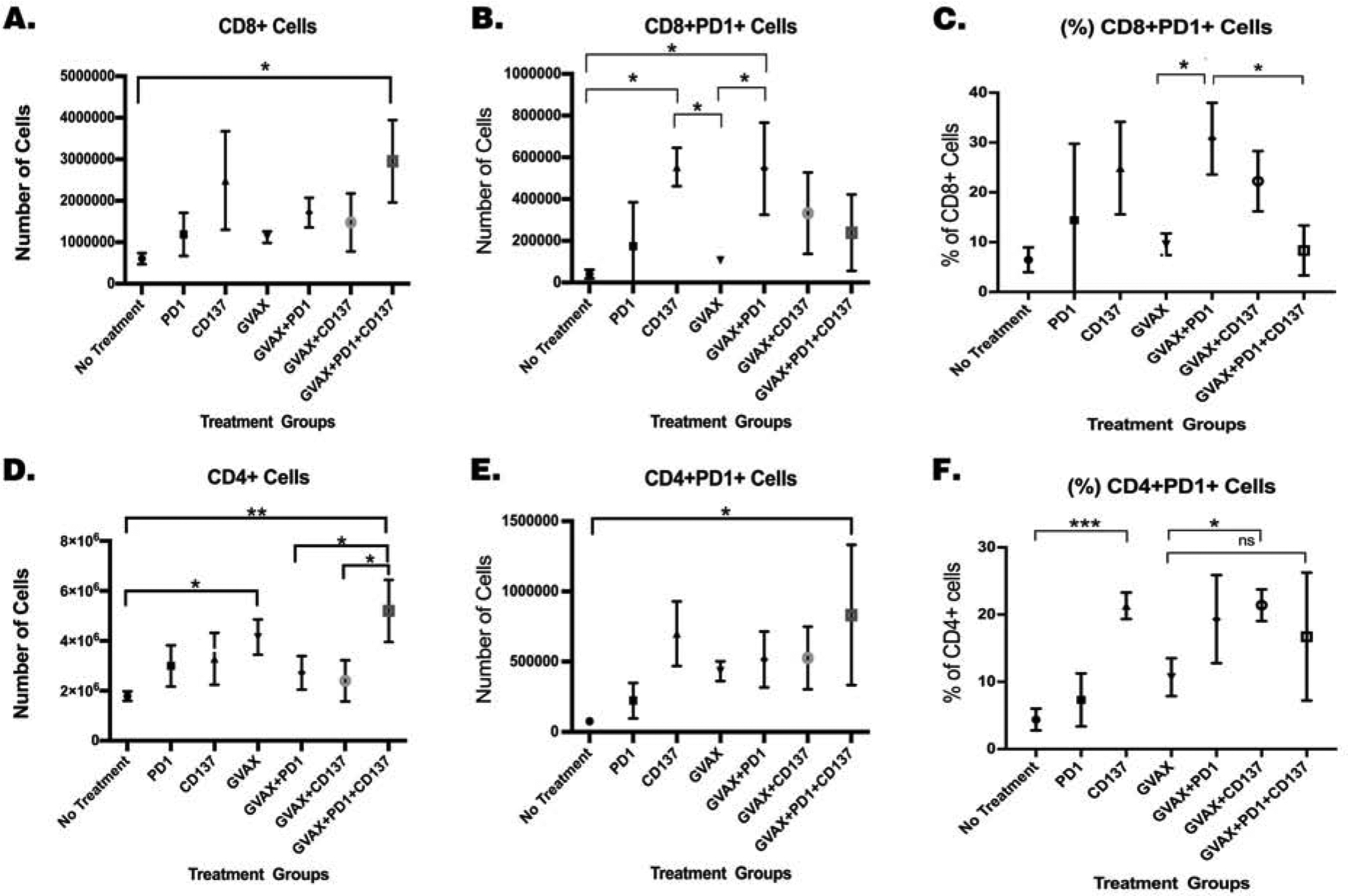
Figure 3. Treatment with GVAX, αPD-1 and αCD137 increased the number of CD4+ and CD8+ cells within the tumor, along with decreased PD-1 expression on CD8+ T cells.

Mice were inoculated with 2×10⁵ KPC cells via the hemispleen surgery, treated and then sacrificed 14 days after KPC cell inoculation for isolation of tumor-infiltrating immune cells for flow cytometry analysis. Normalized numbers of (A.) CD8+ and (D.) CD4+ T cells. Normalized numbers of (B.) CD8+PD-1+ and (E.) CD4+PD-1+ T cells. Percentages of (C.) CD8+PD-1+ and (F.) CD4+PD-1+ T cells among CD8 cells and CD4 cells, respectively. Unpaired t tests were done. * p<0.05; ** p<0.01; *** p<0.001; ns, not significant. Numbers of T cells were normalized by the total cell counts of the single cell suspension of the livers. Data represents mean ±SEM in one representative experiment (n=3 per treatment group per experiment, repeated twice).
