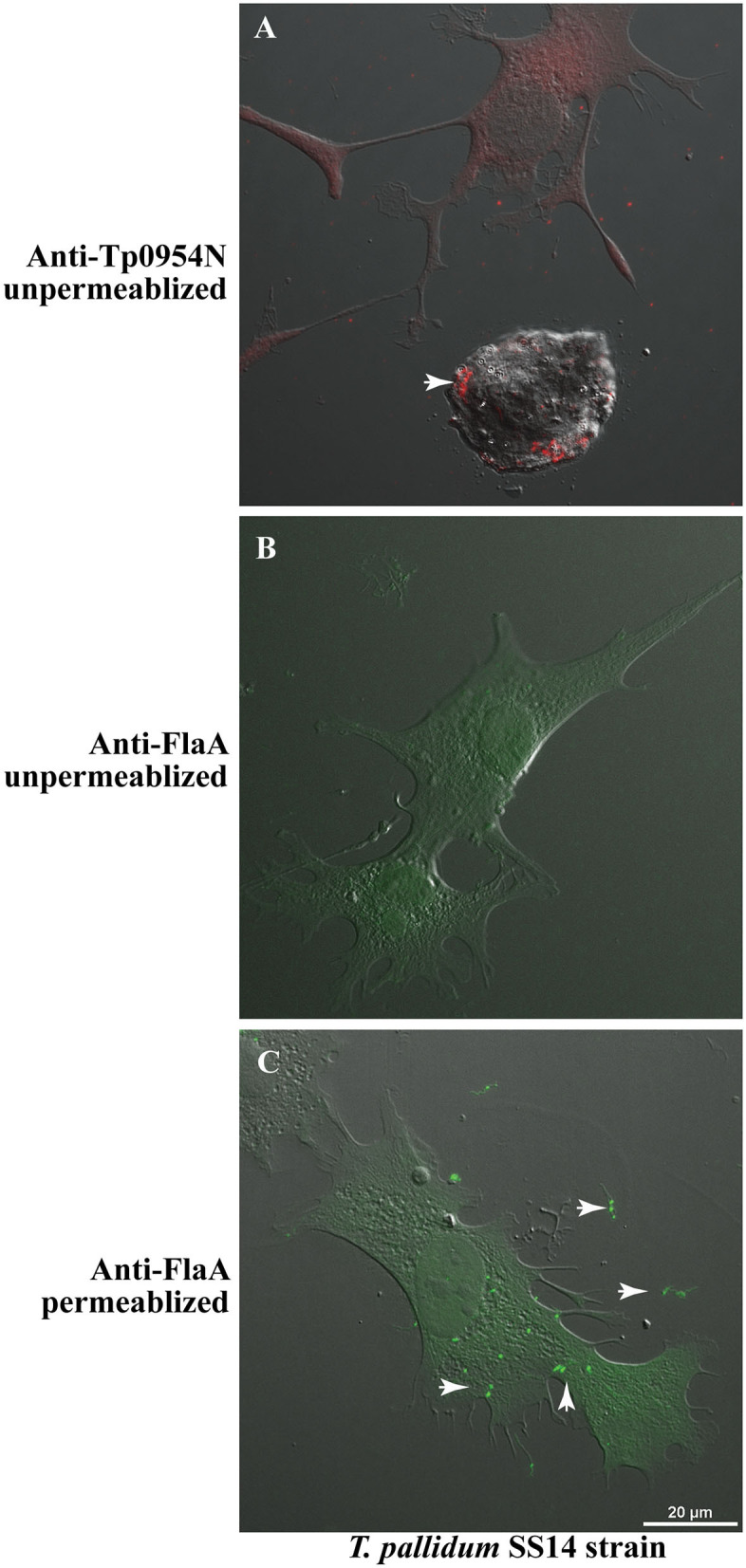
Figure 6.

IFA of T. pallidum co-cultured with low passage rabbit epithelial Sf1Ep cells. (A) IFA using primary polyclonal mouse anti-Tp0954N antibodies followed by TRITC-conjugated secondary antibodies using unpermeabilized T. pallidum SS14 strain co-cultured with Sf1Ep cells showed punctate staining confirming the surface localization of Tp0954. (B) The lack of staining of flagella by anti-FlaA in unpermeabilized T. pallidum indicate that integrity of outer membrane spirochetes was maintained during IFA procedure, and (C) staining of flagellar protein after permeabilization indicate antibodies reactivity and periplasmic location of flagella. Bar indicates 20 μ size.
