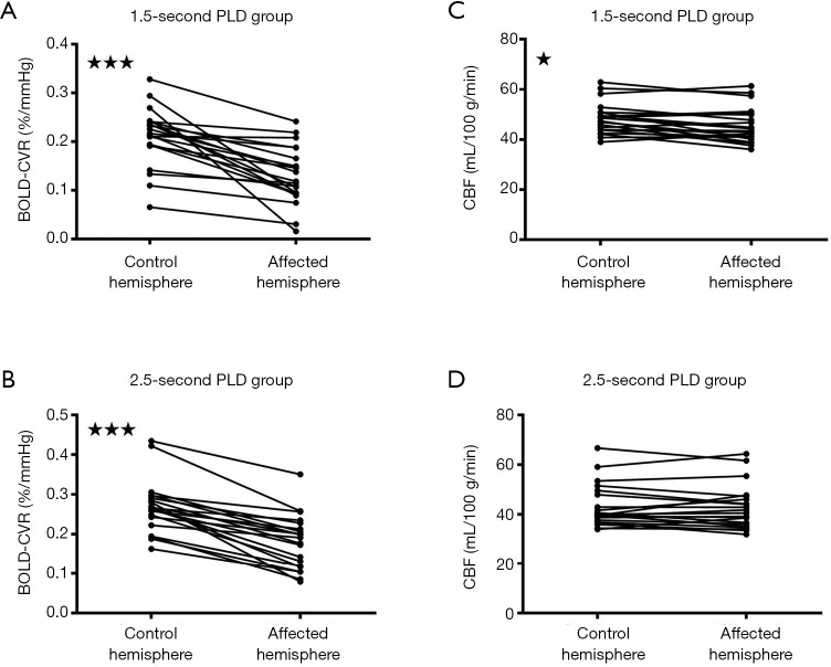
Figure 3.
Figures showing different post-labeling delays (PLDs) and their effects on cerebral blood flow (CBF) when blood oxygen level dependent cerebrovascular reactivity (BOLD CVR) was impaired, according to the hemisphere-based analysis. When BOLD CVR values were significantly decreased (A,B), CBF values were significantly correspondingly decreased for the 1.5-s PLD arterial spin labeling (ASL) protocol (C), although less pronounced compared to the region of interest (ROI) analysis. Such a difference was not observed for the 2.5-s PLD ASL protocol (D). *, P<0.05; ***, P<0.0001.

