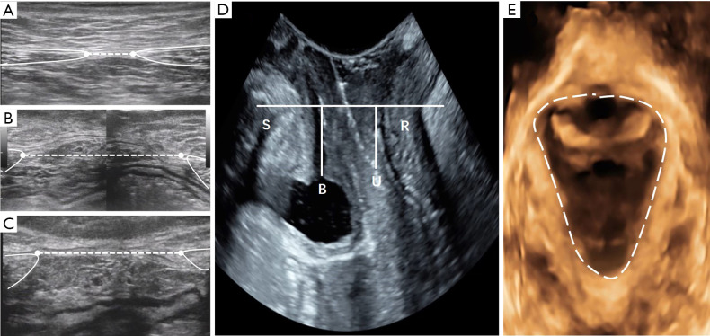
Figure 1.
(A-C) Ultrasonographic images of the linea alba and the rectus muscles in a 20-year-old woman with a BMI of 20.5 kg/m2, G1P1. (A) Transverse scan showing an IRD of 10 mm at 3 cm I-U; (B) 43 mm at the umbilicus; (C) 36 mm at 3 cm S-U; (D) the bladder neck was 12 mm above the line of reference, and the lowest point of the cervix was 10 mm above the line of reference. No significant degree of posterior compartment prolapse was observed; (E) the area of levator hiatus (dotted line) was 16 cm2. Note the distorted linea alba in (B,C). G1P1, first pregnancy; S, symphysis pubis; B, bladder; U, uterine; R, rectal ampulla, IRD, inter-rectus distance; I-U, infraumbilical; S-U, supraumbilical.

