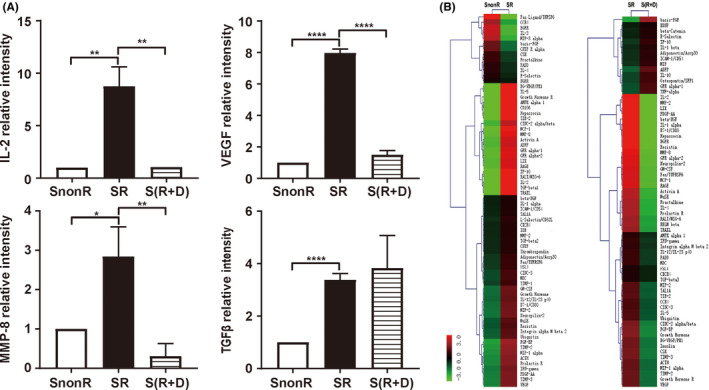
Figure 4.

Increase in inflammation‐ and metastasis‐related factors released following SR. A, Relative expression of interleukin‐2 (IL‐2), vascular endothelial growth factor (VEGF), matrix metalloproteinase‐8 (MMP‐8), and transforming growth factor‐beta‐1 (TGF‐β1) in conditioned medium from irradiated (SR), irradiated plus drug (celecoxib)‐treated S(R + D), and nonirradiated (SnonR) supernatant based on rat cytokine arrays. Error bars indicate the mean ± SD. B, Hierarchical clustering analysis of all expressed SnonR, SR, and S(R + D). Maps on the left are based on the expression values in SnonR and SR. Maps on the right correspond to the expression values in SR and S(R + D). The expression values are depicted in line with the color scale. The intensity increases from green to red. Green represents all cytokines expressed in SnonR and SR with a fold change < 0.66, and red indicates all cytokines expressed in SR and S(R + D) with a fold change > 1.5. *P < .05, **P < .01, ***P < .001, ****P < .0001
