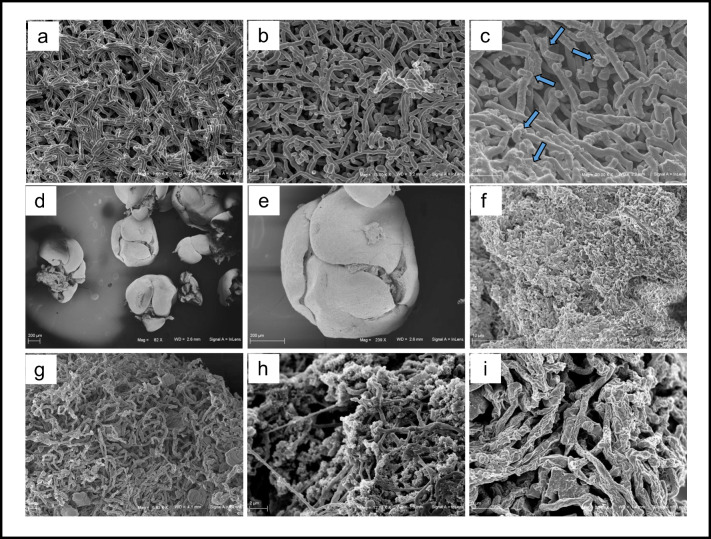
Fig. 2.
SEM pictures of Streptomyces roseochromogenes ATCC 13400 in liquid cultures during hydrocortisone bioconversion at 0, 4, and 48 h, respectively (a–c): the accumulation of material on the cell surface is due to the process of steroid bioconversion and it is indicated by the arrows (c); SEM pictures of Streptomyces roseochromogenes ATCC 13400 whole cells as immobilized in calcium alginate beads (d–f): a tight mycelium was visible in the picture of bead internal view (f); SEM pictures of Streptomyces cyanogriseus ATCC 27426 in liquid cultures during secondary metabolite production at 96, 168, and 264 h, respectively (g–i): morphology changes with increase of cell tangling, branching, and rugosity are visible. (Mag from × 82 to × 20,000, scale bar from 2 to 200 μm). (Preparation of the samples for SEM analyses: small volumes of culture (1 mL) were pelleted end suspended in 4% formalin in PBS for 18 h, dehydrated in increasing ethanol concentrations (from 30 to 100% for 5–15 min), dried in a critical point dryer, and sputtered with platinum-palladium (sputter coater Denton Vacuum Desk V). Fe-SEM Supra 40 Zeiss (5 kV, detector InLens) and Smart SEM Zeiss software were used for observation

