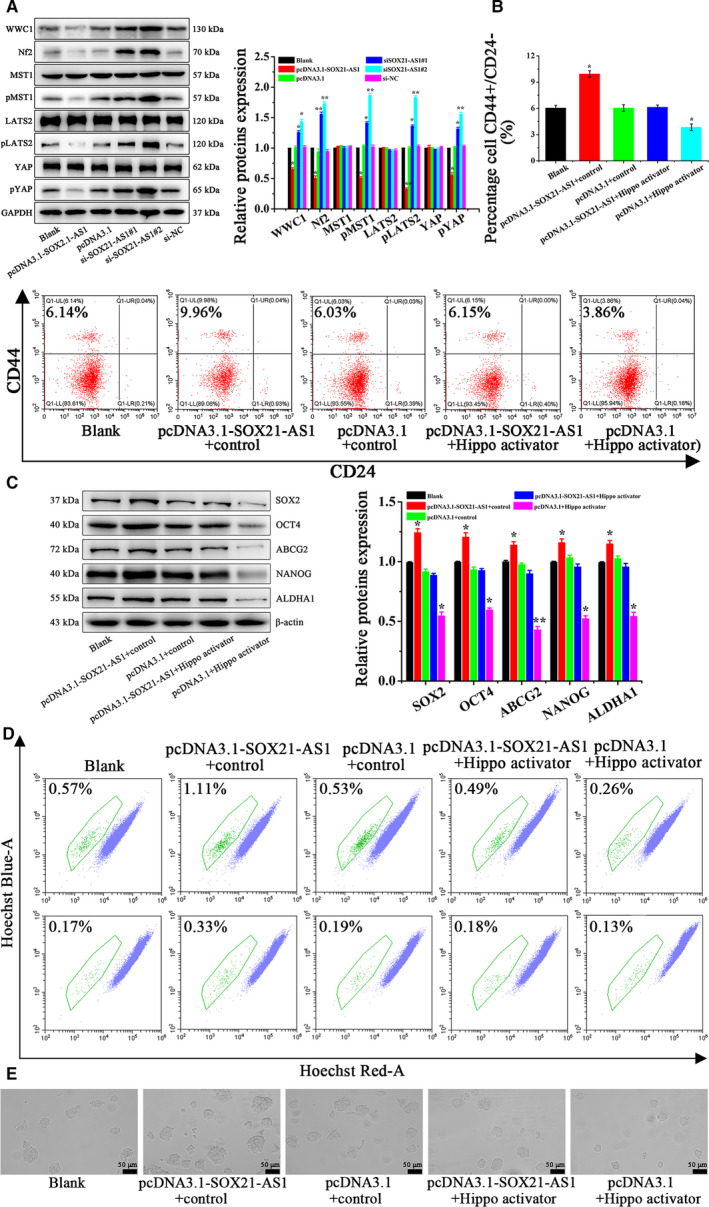
Fig. 4.

(A) The expression of the Hippo signal pathway‐related proteins in differently treated CSC‐MCF‐7 cells (Blank, pcDNA3.1‐SOX21‐AS1, pcDNA3.1, si‐SOX21‐AS1#1, si‐SOX21‐AS1#2, si‐NC) was detected by western blot analysis. (B) The ratio of CD44+/CD24–in CSC‐MCF‐7 cells after different treatments (Blank, pcDNA3.1‐SOX21‐AS1 + control, pcDNA3.1 + control, pcDNA3.1‐SOX21‐AS1 + Hippo activator, pcDNA3.1 + Hippo activator) was detected by flow cytometer analysis. (C) The expression of CSC‐MCF‐7 cell stemness‐related proteins after different treatments (Blank, pcDNA3.1‐SOX21‐AS1 + control, pcDNA3.1 + control, pcDNA3.1‐SOX21‐AS1 + Hippo activator, pcDNA3.1 + Hippo activator) was detected by western blot analysis. (D) The proportion of SP in CSC‐MCF‐7 cells after different treatments (Blank, pcDNA3.1‐SOX21‐AS1 + control, pcDNA3.1 + control, pcDNA3.1‐SOX21‐AS1 + Hippo activator, pcDNA3.1 + Hippo activator) was detected by flow cytometer analysis. (E) Sphere formation ability in CSC‐MCF‐7 cells after different treatments (Blank, pcDNA3.1‐SOX21‐AS1 + control, pcDNA3.1 + control, pcDNA3.1‐SOX21‐AS1 + Hippo activator, pcDNA3.1 + Hippo activator) was detected using a stereomicroscope. Scale bars = 50 μm (n = 3). The results are reported as the mean ± SD of three experiments. *P < 0.05; **P < 0.01 (Student′st‐test).
