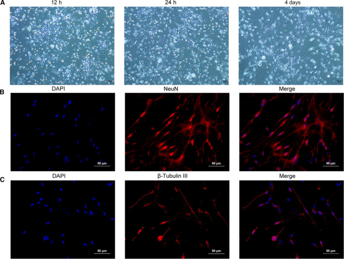
Fig 1.

Isolation and culture of rat trigeminal ganglion neurons. (A) Isolated rat trigeminal ganglion neurons were cultured for 12 h, 24 h and 4 days, and then images were acquired by inverted microscope. (B) Isolated rat trigeminal ganglion neurons were stained with the nuclear dye DAPI (blue) and immunostained for NeuN (red). (C) Isolated rat trigeminal ganglion neurons were stained with β‐Tubulin III. Each experiment was repeated three times with similar results. Scale bars: 50 μm.
