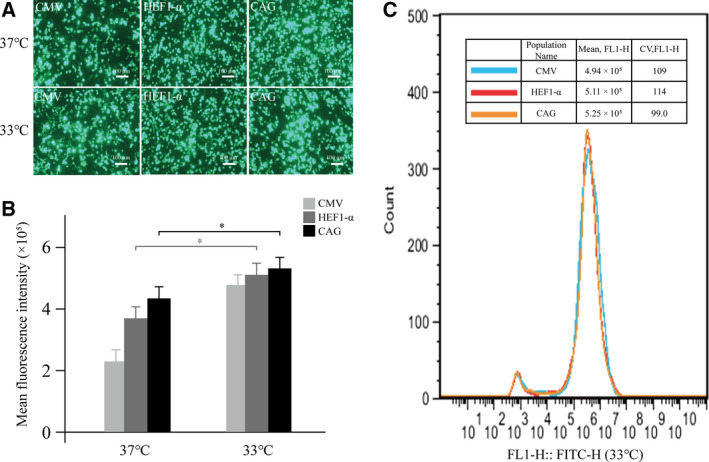
Fig. 5.

Effects of mild hypothermia on the expression of eGFP under different conditions. (A) Cells were transferred from 37 to 33 °C for 24 h and cultured for 7 days post‐transfection. The fluorescence intensity was observed under a fluorescence microscope, scale bars: 100 μm. (B) Cells were transferred from 37 to 33 °C for 7 days at 12, 24, and 48 h post‐transfection, and the expression levels of eGFP were detected by flow cytometry. (C) The eGFP MFI was determined by flow cytometry in a stably transfected cell pool with three highly expressed promoters, CAG, HEF‐α, and CMV mutant. The culture temperature was transferred from 37 to 33 °C 24 h after transfection. Cells were collected after 30 days, and eGFP fluorescence was measured by FACSCalibur. All the experiments were repeated three times. SEM is indicated (Student’s t‐test, *P < 0.05).
