Fig. 1.

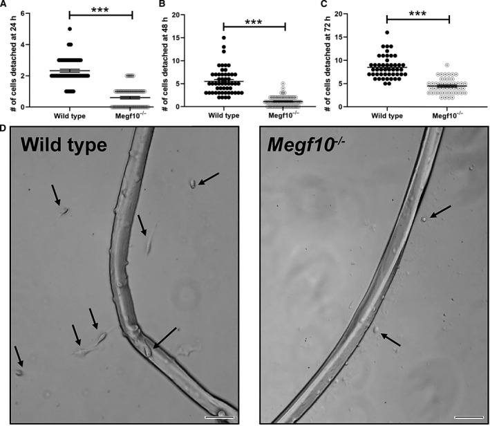
(A) Single intact live myofibers were isolated from wild‐type and Megf10−/− mice and cultured in serum‐rich medium in glass chamber slides. Detached satellite cells were counted for each myofiber at (A) 24, (B) 48, and (C) 72 h timepoints. Each dot represents the satellite cell count for one myofiber. The number of myofibers evaluated were: (A) wild‐type, n = 70 myofibers; Megf10−/−, n = 70 myofibers; (B) wild‐type, n = 52 myofibers; Megf10−/−, n = 70 myofibers; (C) wild‐type, n = 47 myofibers; Megf10−/−, n = 47 myofibers. Comparisons between wild‐type and Megf10−/− myofiber counts were performed at each time point by unpaired t‐tests; ***, P < 0.001. (D) Representative images are shown at 72 h. Arrows indicate satellite cells that have detached and migrated from the isolated myofibers. Scale bar, 50 μm.
