-
A
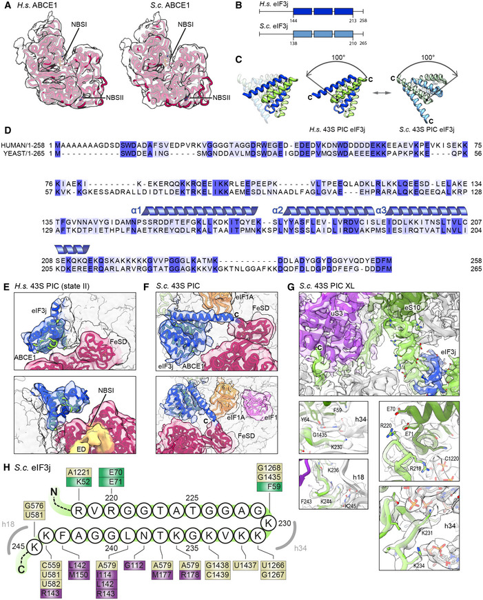
Model for ABCE1 fit into low‐pass‐filtered density to demonstrate the hybrid semi‐open/closed conformation of ABCE1 in native yeast and human 43S PICs.
-
B
Schemes of H.s. and S.c. eIF3j indicating the length of N‐ and C‐termini; the three helices present in the crystal structure of human eIF3j (PDB 3BPJ) are indicated.
-
C
View highlighting the rearrangement (100‐degree rotation) of eIF3 in the human (eIF1A‐lacking) and yeast (eIF1A‐containing) 43S PICs.
-
D
Alignment between H. s. and S. c. eIF3j shows 24.6% identity and 53.1% similarity for the full‐length protein. Dark blue boxes indicate conservation, light blue boxes indicate similarity. For the sequence (three α‐helices) present in the human X‐ray structure (PDB 3BPJ from residues 144‐213 in protomer 1 and 144‐216 in protomer 2), identity/similarity is 32.4%/66.2%, corroborating the reliability of the yeast homology model.
-
E, F
Fits of the human eIF3j crystal structure and the yeast homology model into the corresponding density.
-
G
Interactions of the eIF3j C‐terminus (protomer 2) with the 40S head and body shown with models fit into the cryo‐EM map of the crosslinked yeast 43S‐PIC.
-
H
Schematic representation summarizing interactions of the eIF3j C‐terminus with the 40S. Colored boxes indicate residues interacting with eIF3j, with brown representing 18S rRNA, purple representing uS3, and green representing eS10.

