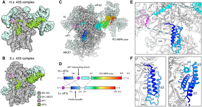
Figure 6. Arrangement of the eIF3c‐NTD in human and yeast 43S PICs.

- Cryo‐EM map obtained after focused sorting of the human 43S PIC on the TC: when low‐pass filtered at 6 Å, it shows the density of almost complete eIF3c‐NTD in the ISS.
- Cryo‐EM map of the yeast 43S PIC low‐pass filtered at 6 Å.
- Model for human eIF3c in the TC‐containing 43S colored in rainbow (C) and scheme of the alignment between human and yeast eIF3c sequences, colored accordingly (D). The eIF1‐interacting stretch present in the N‐terminus of S.c. eIF3c shows 32.0/56.0% sequence identity/similarity with an insert C‐terminal of the conserved 4‐helix bundle conserved in mammals.
- Zoomed view highlighting the position of the eIF3c NTD and eIF1 in the 40S ISS.
- Molecular model for the 4‐helix bundle interacting with 40S rRNA and r‐proteins.
- Molecular model for the 4‐helix bundle interacting with 40S rRNA and r‐proteins.
