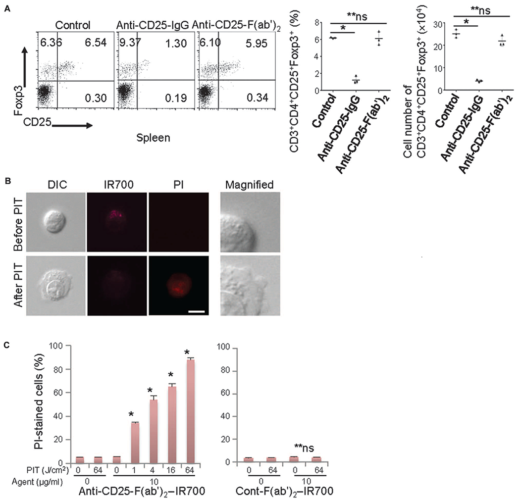
Fig. 1. Anti-CD25-F(ab’)2 lacks Treg depletion effect, but NIR-PIT with anti-CD25-F(ab’)2–IR700 kills target cells.

(A) Intravenously injected anti-CD25-IgG (100 μg) systemically depleted CD4+CD25+Foxp3+ Tregs, but anti-CD25-F(ab’)2 (100 μg) did not significantly deplete these cells within the CD4 T cell population 1 day after injection (n = 3) [*P < 0.0001, one-way analysis of variance (ANOVA) with Dunnett’s test; **P = not significant (ns)]. (B) HT-2-A5E cells (mouse T lymphocytes) incubated with anti-CD25-F(ab’)2–IR700 for 6 hours were examined under a microscope before and 0.5 hour after NIR light irradiation (4 J/cm2). NIR light irradiation induced cellular swelling, bleb formation, and necrosis of the cells, as indicated with propidium iodide (PI) staining (scale bar, 10 μm; right magnified view, ×4). DIC, differential interference contrast. (C) Cell necrosis induced by NIR-PIT increased in a NIR light dose–dependent manner, as determined by flow cytometry analysis with PI staining (left graph, n = 3; *P < 0.0001 versus 0 J/cm2, unpaired t test). No significant cell killing was detected when a control-F(ab’)2–IR700 was used (right graph, n = 3; **P = ns versus 0 J/cm2, unpaired t test).
