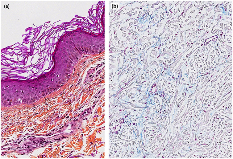
Figure 2.
Skin histology. (a) A hematoxylin phloxine saffron–stained section at 20× magnification showing rare necrotic keratinocytes with discrete vacuolization of the basal cell layer at the basement membrane zone and perivascular lymphocytic infiltrates and (b) staining with blue Alcian (pH 2.5) at 10× magnification highlighting increased dermal mucin deposition.

