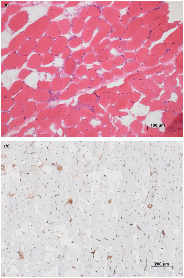
Figure 3.

Muscle histology. (a) Hematoxylin and eosin section of semimembranosus muscle biopsy showing scattered purple staining necrotic and regenerative fibers without lymphocytic infiltration (200× magnification) and (b) immunohistochemical preparation for MHC-1 showing overexpression restricted to scattered necrotic fibers and lack of capillary dropout (100× magnification).
MHC: major histocompatibility complex.
