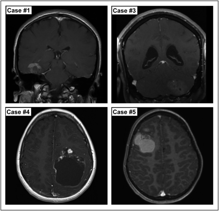
Figure 1.

Representative pre‐operative imaging from four cases of intracranial DSRCTs, showing T1 weighted MRIs with contrast. Images demonstrate the variety of radiologic findings including supratentorial and infratentorial locations, the variable degree of a cystic component and relative degree of enhancement. Case #1: A right temporal heterogeneously enhancing mass. Case #3: A left cerebellar heterogeneous minimally enhancing mass. Case #4: A left parietal cystic mass with enhancing nodules. Case #5: A right frontal intrinsically T1 hyperintense mass with only focal nodular enhancement.
