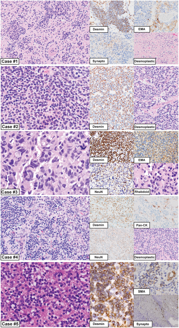
Figure 2.

Morphologic appearance of intracranial DSRCTs for cases 1–5. Case #1: Regions of this tumor resembled an astroblastoma‐like glial neoplasm, while in other areas there was a markedly desmoplastic stroma with interspersed small round cells. Immunostaining for desmin, EMA and synaptophysin are shown. Case #2: This tumor had a uniform solid appearance with sheets of hyperchromatic nuclei, and only focal areas of mild desmoplasia. Desmin was strong and diffusely positive. Case #3: The histology of this case resembled an anaplastic medulloblastoma, including cell‐wrapping, large cells and nuclear molding. Rhabdoid features were appreciated focally; however, a desmoplastic stroma was not present in this case. Immunostaining for desmin, EMA and NeuN are shown, depicting the globular desmin staining that is often described for DSRCTs. This was the only case in our series with extensive epithelial marker expression. Case #4: This tumor appeared low grade with prominent areas of desmoplasia. Immunostaining for desmin, pan‐cytokeratin and NeuN are shown. Case #5: These tumor cells contained small round blue nuclei, and there were focal areas of desmoplasia. Immunostaining for desmin, EMA and synaptophysin are shown.
