Fig. 4.
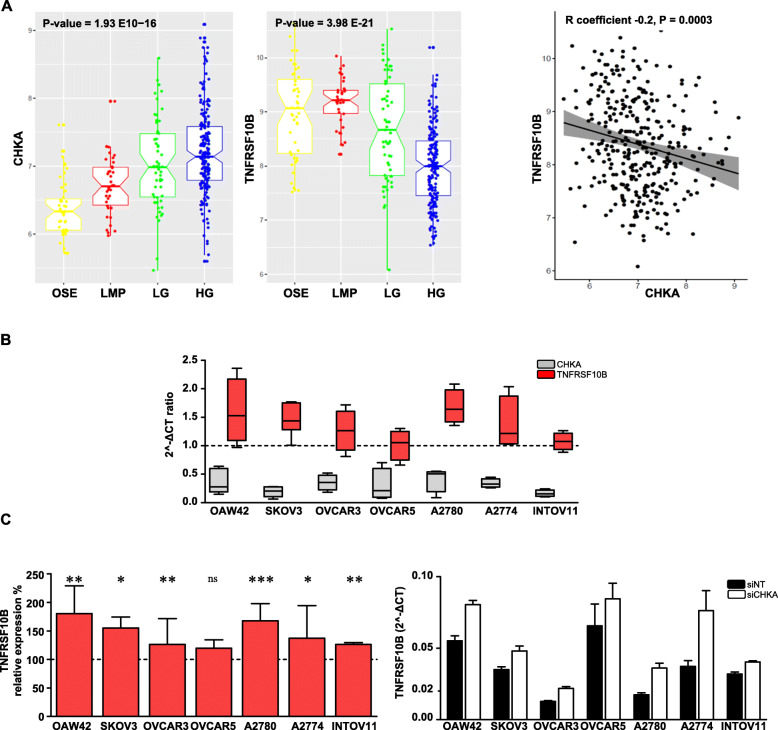
CHKA and TNFRSF10B expression are inversely related in OC samples and cell lines. a. Left panels: Notched box plots showing the log2 expression values of CHKA and TNFRSF10B within preparations of normal ovarian surface epithelium (OSE), low malignant potential (LMP), low grade (LG), high grade (HG) tumors. The boxes represent the interquartile range; the notches the 95% confidence intervals of the median; the whiskers the ranges within 1.5× interquartile range of the upper or lower quartile. Right panel: Inverse correlation of log2 expression between CHKA and TNFRSF10B in the meta-analysis, 95% confidence area (grey region). b. Comparative analysis of CHKA (red boxes) and TNSFRSF10B (grey boxes) transcripts variation in OC cell lines following siCHKA. For each transcript, the relative change as compared to siNT cells is shown. Dotted line (=1) represents no transcript expression variation between siCHKA and siNT samples. c. Relative quantification of TNFRSF10B transcript expression reported as percentage of 2^-ΔCT siCHKA/siNT ratio (left panel); asterisks are referred to a statistically significant differences; a representative experiment is shown (right panel)

