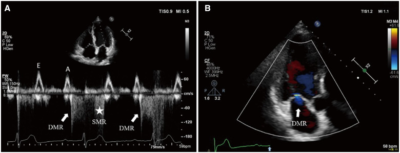
Figure 2.
P-R interval significantly prolongs over 300 ms on the electrocardiogram. Transthoracic echocardiography: (A) PW Doppler of transmitral flow from the apical four-chamber view. After left atrium systole, left ventricular contraction delays and diastolic mitral regurgitation appears with low velocity (solid white arrow) followed immediately by high velocity systolic mitral regurgitation (asterisk). (B) Colour flow Doppler still frame of transmitral flow from the apical three-chamber view. Mild diastolic mitral regurgitation jet (solid white arrow) presents after the sinus P-wave (solid blue arrow on electrocardiogram tracing).

