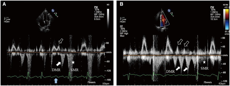
Figure 3.
(A) Atrial flutter with variable conduction on the electrocardiogram. Transthoracic echocardiography: PW Doppler of transmitral flow from the apical four-chamber view. During a 4:1 conduction, the third blocked F-wave (solid blue arrow) drives left atrium contraction accompanied by mitral valve forward flow (hollow white arrow), which is then followed by diastolic mitral regurgitation (solid white arrow) in late diastole. (B) Atrial fibrillation with a 1.5 s interval on the electrocardiogram. Transthoracic echocardiography: Multiple mitral valve forward flows (hollow white arrows) are followed closely by diastolic mitral regurgitations (solid white arrows), respectively until next left ventricular contraction. For the diastolic mitral regurgitation in both situations, the flow velocity is slightly higher than previous mitral valve forward flow.

