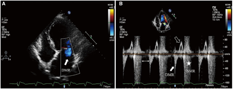
Figure 4.
Transthoracic echocardiography: (A) Colour flow Doppler still frame demonstrates diastolic mitral regurgitation jet (solid white arrow) from the apical four-chamber view after T-wave (solid blue arrow on electrocardiogram tracing). (B) PW Doppler of transmitral flow dictates two mitral regurgitation spectrums at different time of the cardiac cycle, where diastolic mitral regurgitation (solid white arrow) and systolic mitral regurgitation (asterisk) both exist. Transmitral valve forward flow signal only appears in late diastolic phase (hollow white arrow). A special ‘blank period’ existing between the two mitral regurgitation spectrums (double-headed arrow) may be due to dyssynchronous and paradoxical left ventricular movement among segments.

