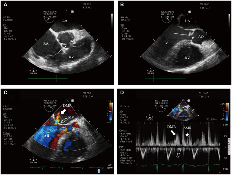
Figure 5.
Transoesophageal echocardiography: (A) Midoesophageal short axis view of the aortic valve demonstrates the morphology of bicuspid aortic valve with minor thickening (arrow). (B) Midoesophageal long axis view reveals vegetations and prolapse of aortic valve (arrow). (C) Colour flow Doppler still frame demonstrates severe aortic regurgitation jet and diastolic mitral regurgitation jet (solid white arrow), and the presystolic closure of the mitral valve (hollow white arrow) before P-wave (solid blue arrow on electrocardiogram tracing). (D) PW Doppler of transmitral flow dictates two mitral regurgitation spectrums at different time of the cardiac cycle, diastolic mitral regurgitation (solid white arrow), and systolic mitral regurgitation (asterisk), and no obvious late diastolic forward flow by left atrium contraction (hollow white arrow). AO, aorta; AR, aortic regurgitation; LA, left atrium; LV, left ventricle; MV, mitral valve; RA, right atrium; RV, right ventricle.

