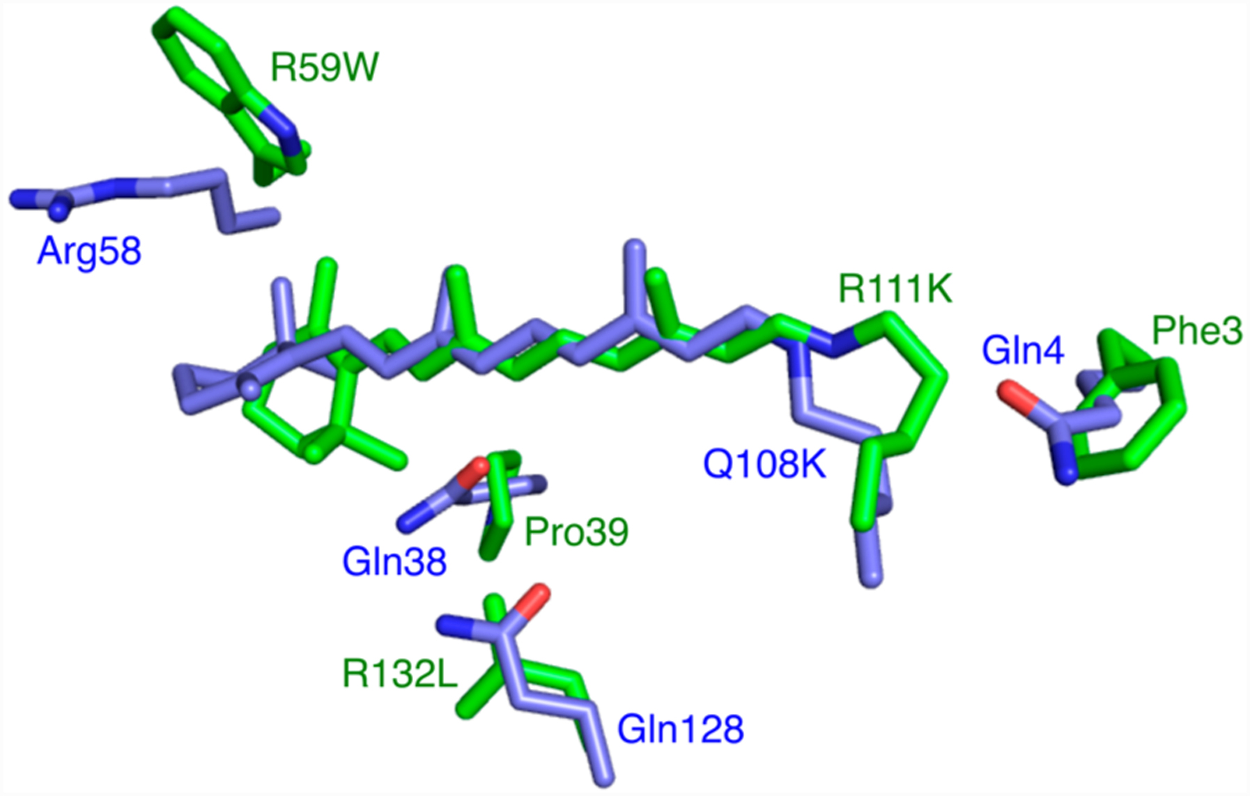
Figure 1.

Structures of the CRABPII mutant R111K:R132L:Y134F:T54V:R59W (green, PDB ID 4I9S) and the hCRBPII mutant Q108K:K40L (blue, PDB ID 4RUU) were overlaid, and the region around the retinal binding site is shown. Note the three glutamines in hCRBPII are substituted with hydrophobic residues in CRABPII, leading to a much higher pKa for the former. Atoms, here and throughout, are colored as follows: O is red, N is blue, and C is green unless otherwise indicated.
