
Figure 4.
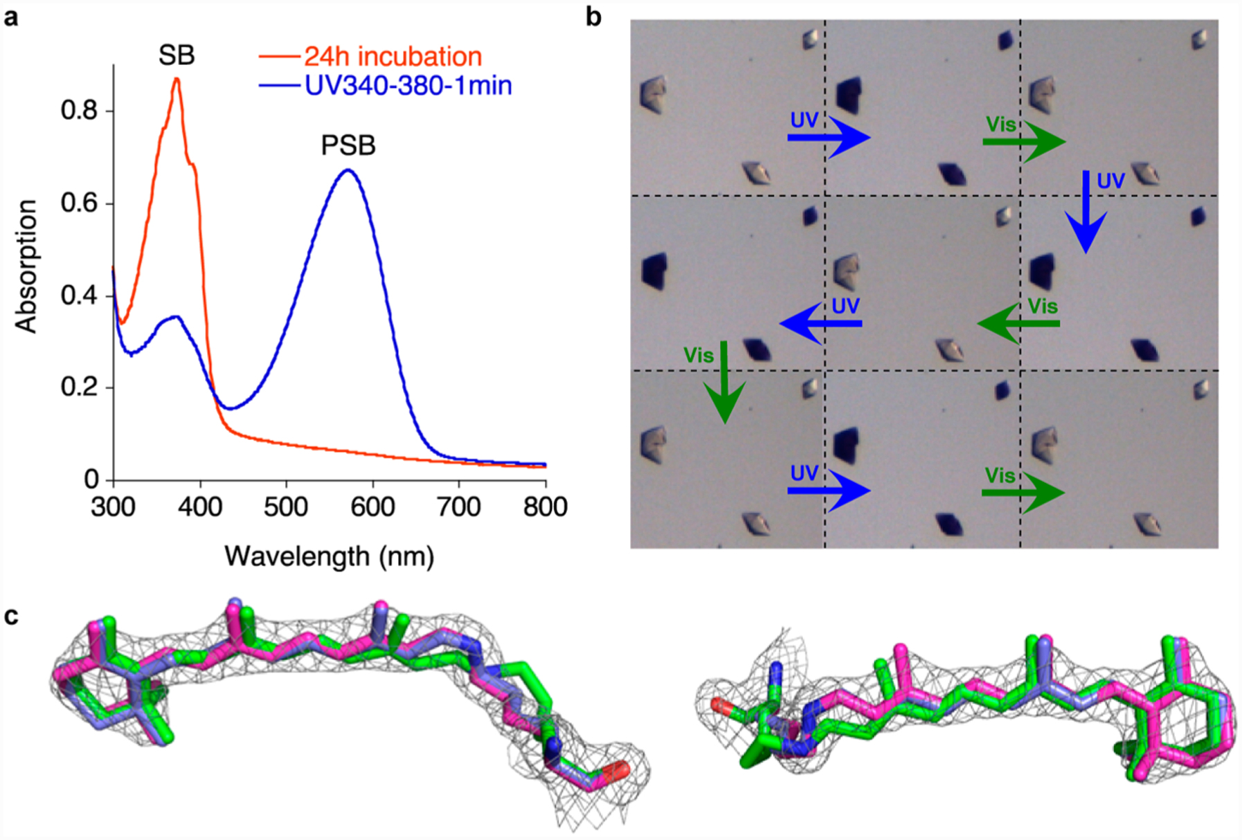
Photointerconversion in both solution and crystalline state. (a) UV irradiation of M2 (UV band-pass filter) after complete PSB loss (24 h incubation, red spectrum) shows the PSB recovery in solution (blue spectrum). (b) Cycles of UV and visible light irradiation of M2 crystals (each cycle is 5 min UV light and 5 min white light irradiation at pH 7.5). (c) The structure of M2 mutant (magenta, PDB ID: 4YFR, 1.95 Å resolution) obtained from a crystal that was UV irradiated for 30 min, overlaid with the metastable cis-PSB structure (blue, PDB ID: 4YFP, see text for description). The electron density was contoured at 1.5 σ; the structure clearly shows the bound retinal adopts a cis imine geometry; also overlaid is the crystal structure of the trans-SB (green, PDB ID: 4YFQ) to highlight the change in structure and its inability to fit the electron density obtained for the UV irradiated crystals. While the left view clearly shows the lysine side chain of the trans imine isomer is out of the density, the right view shows the motion of the retinal.