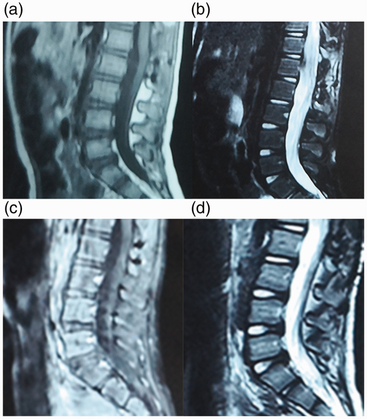
Figure 2.
Representative postoperative magnetic resonance imaging (MRI) scans in a 3-year-old girl that had pain in her thighs for more than 3 months and had difficulty walking when she was admitted for assessment and diagnosis. No tumour recurrence was observed. (a) Sagittal T1 and (b) sagittal T2-weighted MRI images on the third day after surgery; (c) sagittal T1 and (d) sagittal T2-weighted MRI images at 9 months after surgery.

