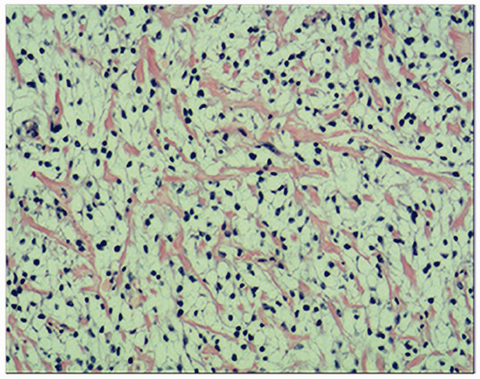
Figure 3.
Representative photomicrograph showing the histopathological findings from a sample of the tumour taken from a 3-year-old girl that had pain in her thighs for more than 3 months and had difficulty walking when she was admitted for assessment and diagnosis. The histology showed sheets of polygonal tumour cells with round or ovoid nuclei and clear cytoplasm. The tissue section was stained with haematoxylin and eosin stain. Scale bar 100 µm. The colour version of this figure is available at: http://imr.sagepub.com.

