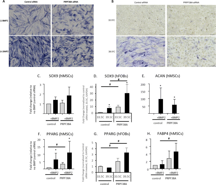
Fig. 5.
PRPF38A knockdown induced a morphological change in two osteoblast cell models. a Cell morphology in hMSCs before (top) and after BMP2 treatment (bottom) with control siRNA (left) and PRPF38A knockdown (right). Representative color bright-field images of a typical alkaline phosphatase stained plate from PRPF38A silenced cells is shown. Similar morphological changes were observed for all three donor lines used in the study. Scale bar, 1000 μm. b PRPF38A knockdown-induced morphological changes were recapitulated in human fetal osteoblasts under permissive growth (33.5 °C; top) and differentiation (39.5 °C; bottom) conditions. Scale bar, 200 μm. c–e Quantitative gene expression of chondrocytic genes SOX9 and ACAN and f–h adipocyte-specific genes PPARG and FABP4. For hMSCs, data is from two technical replicates from three unique donor lines were averaged. For hFOBs, three technical replicates were averaged. Levels of ACAN and FABP4 were undetectable in hFOBs, even in reactions with up to 600 ng of cDNA (twice the amount used for qPCR of other targets). *p < 0.05 comparing non-treated to treated cells (BMP2 or 39.5 °C for hMSCs and hFOBs, respectively) for each siRNA, #p < 0.05 comparing control siRNA to siRNA for gene of interest. Error bars represent standard deviation

