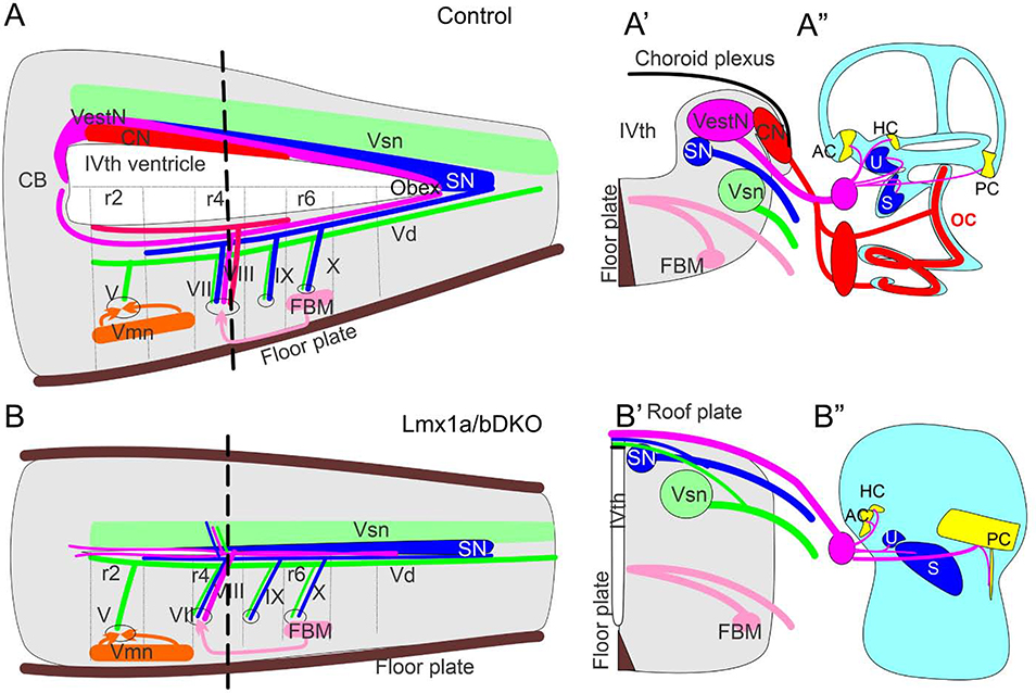
Fig. 10. Simultaneous loss of Lmx1a and Lmx1b disrupts the inner ear, central projections and cochlear/vestibular brainstem nuclei.
(A, A’) Control mice have a large IVth ventricle covered by a choroid plexus. Specific nuclei develop in the alar and basal plate of the hindbrain. CN – cochlear nuclei, which develop in r2–5, VestN – vestibular nuclei, which extend from the caudal cerebellar vermis to the obex, SN – solitary tract nuclei, Vsn – viscerosensory (trigeminal) nuclei, FBM – facial branchial motor neurons, Vmn – ventral motor neurons. Each set of nuclei receives distinct sensory afferents. For example, auditory information is provided by spiral ganglion neuronal projections to cochlear nuclei.
(B, B’) In Lmx1a/b DKO mice, excitatory neurons of cochlear and vestibular nuclei do not develop. Segregated projections to viscerosensory trigeminal nuclei (Vsn) and solitary tract nuclei (SN) were detected in these mice. Inner ear vestibular afferents project to and cross the roof plate in r4, with limited caudal extension. Similar to inner ear afferents, some solitary tract and trigeminal fibers project to the roof plate. In contrast, basal plate development and innervation (motor neurons) were not significantly affected in Lmx1a/b DKO mice.
(A”, B”) Organ of Corti (OC), spiral ganglion (red oval near the ear in control mice) and spiral ganglion projections were not detected in Lmx1a/b DKO mice. In contrast, although altered in size or shape, vestibular inner ear components (AC, HC, U, S, PC and vetibular ganglion - pink circle near the ear) still develop in Lmx1a/b DKO embryos.

