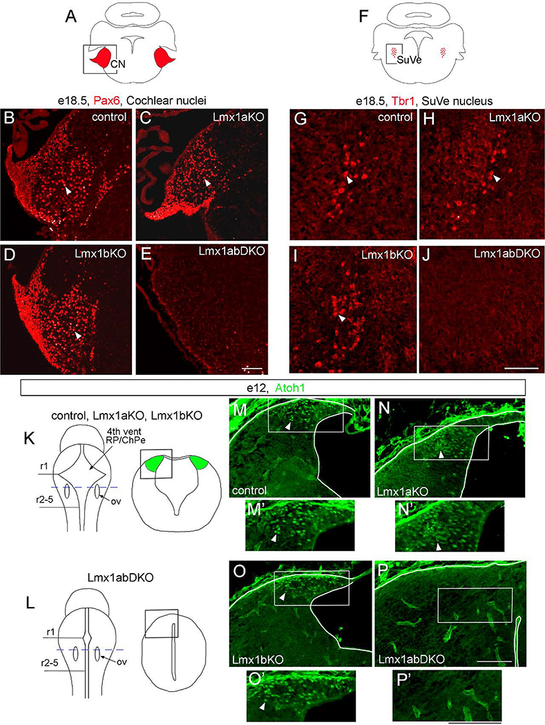
Fig. 9. Simultaneous, but not individual, loss of Lmx1a and Lmx1b prevents the development of excitatory neurons in cochlear and vestibular nuclei.
Pax6 (B-E), Tbr1 (G-J), and Atoh1 (M-P’) immunostained transverse sections of the hindbrain at indicated stages. High magnification panels correspond to regions boxed in adjacent diagrams (A, F, K, L) or low magnification panels.
(A-E) Numerous Pax6+ neurons were present in the cochlear nuclei (CN) of wild type control, Lmx1a KO, and Lmx1b KO mutants (arrowheads) but were not detected in Lmx1a/b DKO embryos.
(F-J) Tbr1+ neurons were present in the superior vestibular nuclei (SuVe) of wild type control, Lmx1a KO, and Lmx1b KO mutants (arrowheads) but were not detected in Lmx1a/b DKO embryos.
(K, L) Diagrams illustrating location of sections showing lower RL in control and Lmx1a/b mutant embryos (M-P). Sections were taken at the level of otic vesicles (ov, blue dashed line in whole mount diagrams in K, L). Tissue morphology is illustrated in right diagrams in K and L. (M-P’) Numerous Atoh1+ progenitors were present in the lower RL of wild type control, Lmx1a KO and Lmx1b KO mutants (arrowheads) but not in Lmx1a/b DKO embryos.
Scale bar: 100 μm.

