Fig. 3.
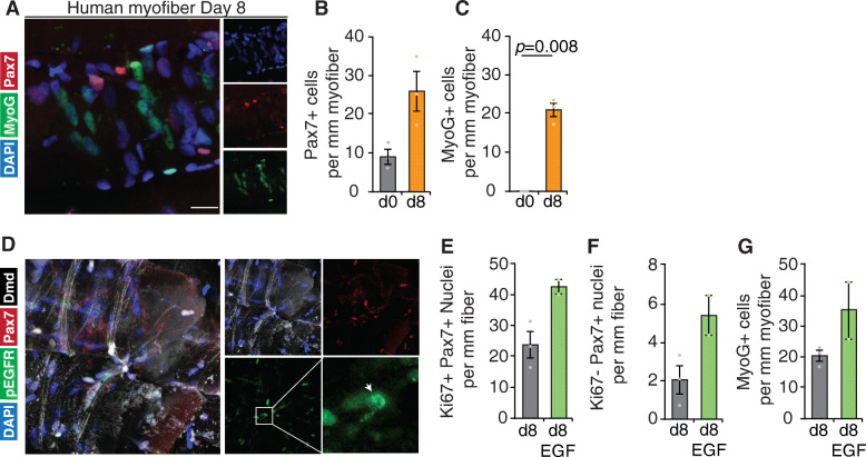
Human satellite cell expansion and differentiation can be tuned in situ. a Representative image (magnification from Figure S3A) of human satellite cells and Myogenin (MyoG) expressing differentiating progenitors cultured on 8-day human Psoas minor myofiber cultures stained with DAPI (blue), MyoG (green), and Pax7 (red). Quantification of b number of Pax7-expressing cells per millimeter of myofiber, c MyoG-expressing cells per millimeter of myofiber. d Representative images of human satellite cells expressing phosphorylated active EGFR (p-EGFR) stained with DAPI (blue), p-EGFR (green), Pax7 (red), and dystrophin (white). White arrow denotes localized p-EGFR expression. Quantification of e satellite cells expressing Ki67 per millimeter of myofiber and f Ki67-negative satellite cells per millimeter of myofiber following EGF treatment or vehicle control of human myofibers. g Quantification of number of MyoG-expressing cells per millimeter of myofiber following EGF treatment of human myofibers. b, c, e–g Error bars represent means ± SD (EGF) and means ± SEM (control); b, c n = 3 biological replicates. d–g n = 3 biological replicates control, n = 2 biological replicates EGF

