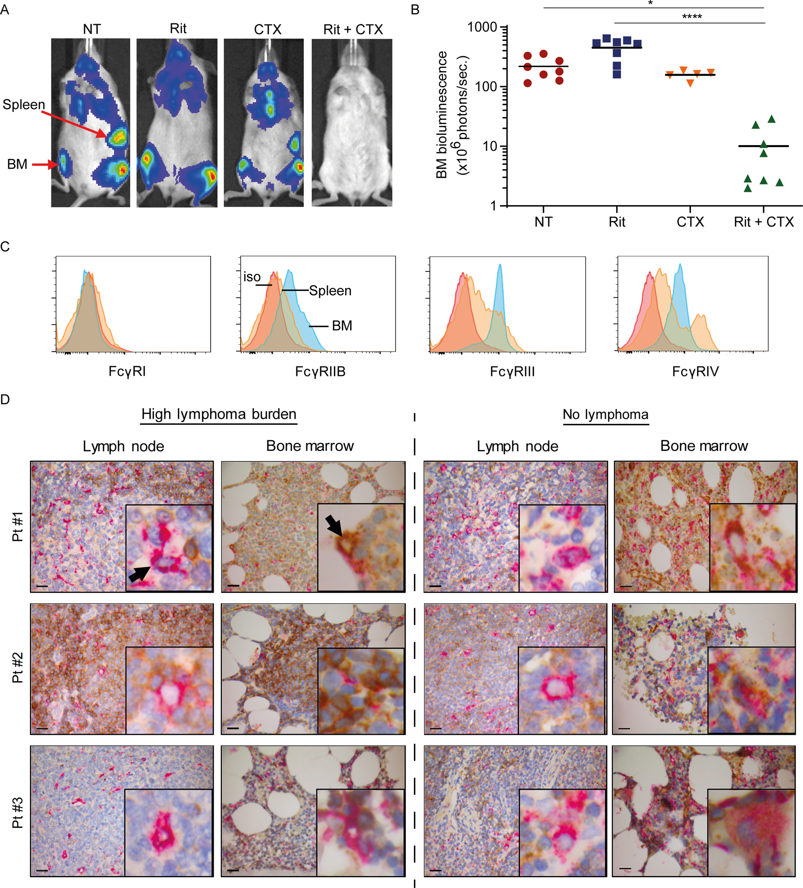
Figure 1. BM niche is resistant to mAb therapy and promotes immunosuppressive effector macrophages.

Ten million luciferase+GFP+ CD20+ human B-cell lymphoma cells were intravenously injected into adult NSG mice (5–8 mice/group from 1 experiment) and were left untreated (NT) or treated with either rituximab (Rit, 10 mg/kg) or CTX (100 mg/kg) as single agents or in combination (Rit+CTX) on day 21 after tumor injection. Tumor burden, indicated by bioluminescence intensity, was monitored on day 21 (before treatment) and 7 days after the indicted treatments. (A) Representative mouse images and (B) bioluminescence signal intensity in the BM. The positions of the spleen and BM are indicated. Each dot represents one mouse.; *p<0.05, ****p<0.0001 by one-way ANOVA. Means are shown. (C) Representative flow cytometry histograms of FcγR expression on F4/80+ macrophages from spleen and BM of a NSG mouse (5–8 mice/group). Red trace: isotype control (iso); orange trace: spleen; blue trace: BM. (D) FFPE diagnostic LN biopsies and staging BM trephines from three consented patients with follicular lymphoma were double-stained with antibodies to human CD68 (pink) and FcγRIIB (brown). Paired LN and BM samples from each patient were mounted on the same slide and stained. Representative IHC images are shown (original magnification x400; scale bar is 20µm) from areas with high lymphoma burden (left panel) and areas with no lymphoma involvement (right panel). The insert in each image represents 10x higher magnification of a representative macrophage taken from the same section. Black arrows point to CD68+ macrophages in the LN and BM.
